Posts

Showing posts from March, 2024

Understanding the Basics of Fat: A Comprehensive Guide

Image
https://educationtechbysapna.blogspot.com/2024/03/lichens-structure-and-function-of.html   The Ultimate Guide to Different Types of Dietary Fats    One of the three macronutrients that give the metabolic system the energy it needs to run properly is fat. Both unsaturated (good fat) and saturated (bad fat) fats are vital components of our daily diet and are required for our continued health. The foods we eat include both unsaturated and saturated fats. Two different kinds of fats:  You must examine the two types of dietary fats—saturated and unsaturated—in greater detail in order to comprehend the function that fats play in a healthy diet. (Trans fats, a third type, are virtually nonexistent in American diet.)   Saturated:  This fat is referred to as "bad" fat. Animal foodstuffs like beef and pork as well as high-fat dairy products like butter, margarine, cream, and cheese are the main sources of it. A lot of quick, processed, and baked items including pizza...

Cilia and Flagella : A comparative analysis

Image
 Cilia and Flagella : A comparative analysis  Although they have similar structures, flagella and cilia are two different kinds of cell organelles that have different lengths and functions. Within each cell, there are hundreds of short and many cilia. Contrarily, there are fewer and longer flagella (often one to eight) per cell. Despite having the same structural makeup, motile cilia and eukaryotic flagella can have different beating patterns. Cell exterior features called flagella and cilia mainly facilitate cell motility. Some cells have a huge number of short, hair-like projections called cilia covering their surface. Only one end of a cell contains the long, thread-like structures called flagella.                Unlike flagella, which beat separately, cilia beat in unison. Only eukaryotic cells contain cilia. Both prokaryotic and eukaryotic cells have flagella. Undulipodia organisms have both motile cilia and flagella. The primary fu...

The Hidden Connections: Surprising Similarities and comparision Between Peroxisomes and Mitochondria

Image
The Hidden Connections:  Surprising Similarities and comparision between Peroxisomes and Mitochondria Peroxisomes and mitochondria are incredibly active types of organelles. They have great plasticity and can take on many forms according on the needs of the cell.  Depending on the physio-pathological environment or the metabolic requirements of the cell, their number and shape can vary. The range size of peroxisomes and mitochondria is 0.1 to 1 μm. On the other hand, their structures are different.  Peroxisomes have a single membrane system encircling them, whereas mitochondria have two membrane systems surrounding them.  Peroxisome and mitochondrial counts in cells differ depending on the type of cell (e.g., mitochondria are plentiful in brown adipose tissue but relatively sparse in white adipocytes).  In terms of fatty acid metabolism, mitochondria degrade the majority of long-chain fatty acids to supply acetyl-CoA for the production of ATP and for anabolic re...

Prions and machanism of transmission of prions

Image
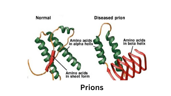
 Prions  The term Prion means proteinaceous infectious particles. Prions are the infectious agents responsible for several neurodegenerative diseases in mammals, like, Creutzfeldt Jakob disease. This happens due to the abnormal folding of the proteins in the brain. It refers to the hypothesis that the infectious agents causing the diseases contain only proteins. It explained why the infectious agents are resistant to ultraviolet radiations. They can break down the nucleic acids, but are receptive to substances that denature proteins. https://educationtechbysapna.blogspot.com/2024/03/how-sem-scanning-electron-microscopy-is.html Prion diseases are transmissible, untreatable, and fatal brain diseases of mammals. Their cause is highly unusual: The host’s normal prion protein can, for unknown reasons, malfunction and assemble into structured aggregates called prions that cause infectious brain disease. This process – which can be underway for years before symptoms appear – likely c...

Glyoxysome and its function

Image
  Glyoxysomes and their purposes  Plant microbodies called glyoxysomes are different from peroxisomes and are involved in certain aerobic pathways in plants, such as fatty acid oxidation and the glyoxylate cycle. In plant cells, lyoxysomes are crucial for seed germination. Unlike peroxisomal enzymes, which emerge later in plant development, active glyoxysome enzymes are present in the microbodies from the very beginning.  https://wwpa.hvldks.com/redirect-zone/ee8dada0 https://educationtechbysapna.blogspot.com/2024/03/lichens-structure-and-function-of.html The primary sources of superoxide and H2O2 generated during the β-oxidation of fatty acids during the catabolism of store lipids in germinating seeds are the enzymes glycolate oxidase and urate oxidase in glyoxysomes. Glyoxysomes appear in the first few days after  seed germination  in  endosperm  cells and associate closely with  lipid  bodies. They disappear after the storage fats are brok...

Secondary structure of protein

Image
 Secondary structure of protein  The term "protein secondary structure" (SS) describes the local configuration of a protein's polypeptide backbone. Pauling13 proposed more than 60 years ago that there are two regular states of supersymmetry (SS): alpha-helix (H) and beta-strand (E), and one irregular form of SS called coil region (C). There are four layers of organization in the structure of proteins. The polypeptide chain's primary structure is its amino acid sequence, which is linear.  https://educationtechbysapna.blogspot.com/2024/03/how-sem-scanning-electron-microscopy-is.html The primary sequence is written according to convention, starting with the amino acid at the N-terminus (which is typically on the left) and ending with the amino acid at the C-terminus. Stretches of adjacent amino acid sequences assume a local conformation that makes up the second level of protein structure, or secondary structure. The two primary forms of secondary structures seen in prote...

The Ultimate Prokaryotic vs. Eukaryotic Cells Comparison

Image
The Ultimate Prokaryotic vs. Eukaryotic Cells Comparison  Prokaryotes are always unicellular, whereas eukaryotes are frequently multicelled organisms; additionally, eukaryotic cells are larger and more complex than prokaryotic cells by an order of magnitude; and finally, eukaryotic DNA is stored in the nucleus of eukaryotes, whereas prokaryotic DNA is stored in the cytoplasm. The Greek words "eu," which means "good," and "karyon," which means "kern," are the roots of the phrase "Eukaryotes," which means "good or true nuclei." Compared to prokaryotes, eukaryotes are larger and more sophisticated.  All the great kingdoms are included, with the exception of Kingdom Monera. Eukaryotes are structurally unique in that their cell walls support and shield the plasma membrane. The plasma membrane, which encloses the cell, regulates the admission and departure of specific molecules. DNA, which is found in the nucleus, is what stores all...

How SEM ( Scanning electron microscopy) is different from TEM (Transmission Electron Microscopy) ?

Image
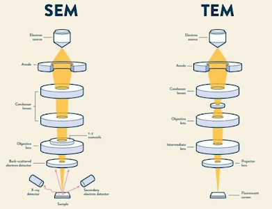
  SEM ( Scanning Electron Microscope) and TEM (Transmission Electron Microscope) A useful technique for obtaining high-resolution images in a range of fields, such as technology, forensics, and biomedical research, is electron microscopy. Compared to optical microscopes, electron microscopes are able to obtain images with significantly higher resolution, which provides information that would not be possible otherwise. Every electron microscope operates by directing a concentrated stream of electrons toward a sample while they are in a vacuum. Similar to how light is used in optical microscopes to form images, interactions between the electron beam and the material produce images. The resulting image provides information on the internal or external makeup of a sample, depending on the kind of electron microscope that is being used. The two most popular forms of electron microscopy are Transmission Electron Microscopy (TEM) and Scanning Electron Microscopy (SEM). The sorts of picture...

Lichens: Structure and Function of Lichens

Image
  Lichens  The word Lichen derives from the Greek Leichen (tree moss, lichen, lichen - like eruption on skin".  A lichen is not a single organism. They are found in a symbiotic, mutually beneficial relationship. The combination of an alga with a fungus creates lichen. The fungus is the main component of lichen.  Lichens  are composed of several organisms from three different kingdoms: fungi, plants, and bacteria.  Lichens are not single organisms but an intimate symbiosis of fungi, most often an ascomycete, and a photosynthetic alga or cyanobacterial species.  The association of these separate organisms is often referred to as a mutualistic relationship, defined as two or more organisms living in a mutually beneficial arrangement.  Most often the symbiosis association includes a second photosynthetic partner (Photobiont) but only one fungus (mycobiont) per lichen.  Spribille  et al . have rewritten this dogma by identifying lichen involv...