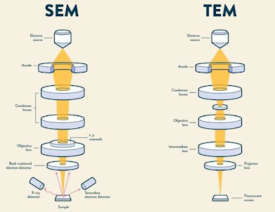
How SEM ( Scanning electron microscopy) is different from TEM (Transmission Electron Microscopy) ?
SEM ( Scanning Electron Microscope) and TEM (Transmission Electron Microscope)
A useful technique for obtaining high-resolution images in a range of fields, such as technology, forensics, and biomedical research, is electron microscopy. Compared to optical microscopes, electron microscopes are able to obtain images with significantly higher resolution, which provides information that would not be possible otherwise.
Every electron microscope operates by directing a concentrated stream of electrons toward a sample while they are in a vacuum. Similar to how light is used in optical microscopes to form images, interactions between the electron beam and the material produce images. The resulting image provides information on the internal or external makeup of a sample, depending on the kind of electron microscope that is being used.
The two most popular forms of electron microscopy are Transmission Electron Microscopy (TEM) and Scanning Electron Microscopy (SEM). The sorts of pictures that can be captured and the ways in which TEM and SEM operate are different. The definition, operation, and comparative analysis of SEM and TEM are covered in this article.
TEM can stand for Transmission Electron Microscopy or Transmission Electron Microscope (TEM) :
How SEM different from the TEM ?
The Science Behind SEM: Mechanism and Function Explored
The SEM scans the specimen back and forth over it with a narrow, tapered electron beam in order to produce an image. Surface atoms are struck by beams that release a tiny shower of electrons known as secondary electrons, which are then captured by a specialized detector. A secondary electron strikes the scintillator, producing a flash of light that is amplified and converted into an electric current by a photomultiplier. A cathode ray tube receives the signal and produces an image similar to a television picture that can be seen or captured on camera.


Comments
Post a Comment